The clinical understanding of ‘dry eye’ diagnosis and need for Lubrizity™️
Conjunctival cul-de-sac
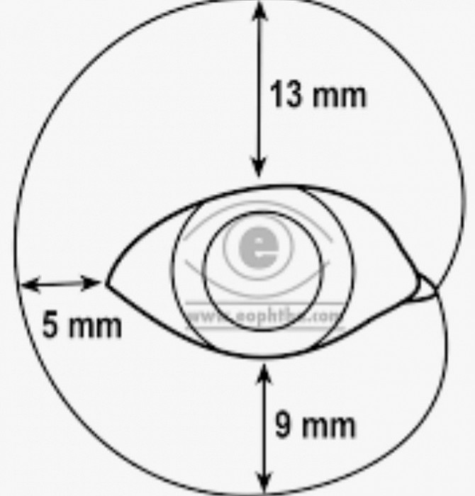
Physiology of the blink
The Tarsal conjunctiva rubs thousands of times a day against the bulbar conjunctiva and cornea. Both have a mucus layer on the epithelium. These layers would stick without an oily lubricant. Tears originate in the lacrimal and accessory glands in the cul-de-sac and pass down under the lids. Mucus membranes stick to each other without oil. The oil from the meibomian glands is postulated to get under the lid by the spherical particles.
The conjunctival surface of the tarsal and bulbar epithelium may be roughened and cause friction. The quality of the oil and meibomian particles must have the proper lubricity to reduce friction. The diagram shows that a large area of the conjunctival sac moves and also needs lubrication.

NEI grading system
Effects of ‘DED’ is Puncatate Stain
The National Eye Institute has done a wonderful job of quantifying punctate stain but we have directed our attention at what causes different patterns on the cornea.

Fundamentals of ocular lubrication
Lubrication is necessary to reduce the friction caused by moving lids as seen in dry eye disease. Below are important considerations:
1. Viscosity and lubricity of the ocular lubricant
2.Rub from lid margins and epithelium on conjunctiva and cornea
3. Flow and distribution of lubrication during blink
Read this link to Tribology for a deeper understanding of lubrication
Direct observation of the precorneal tear film by retro reflection.
It is useful to observe the rising tear film after the blink. It should appear as a mildly viscous film made up of aqueous, oil and microsphericles. Often oil can be seen by birefringence from the filament reflection. If it is missing meibomian particles or appears watery it is abnormal. To learn what it looks like in youth, observe younger patients. Remember, the tear film acts like a lubricant to counter the effects of rubbing from the lids, conjunctiva and ocular rotations. Please be acutely aware that all OTC eye drops only temporarily add to this layer as monitored in the PRECORNEAL tear film. Lubrisity™️, on the other hand, thickens and restores oil and microsphericles for an extended period of time.
See below:
Fluorescein coating
Fluorescein dye temporarily stains the tear film and shows how evenly the tear film is distributed across the ocular surface. The videos after demonstrate dry eye abnormalities.
Normal tear film
Example of corneal punctate stain caused by rubbing from lashes.
Rubbing, poor lubricity of tear film, incomplete tear film wetting by conjunctival scarring, elevation and chalasis are common causes of punctate stain Below are a series of videos demonstrating this.
Rubbing lashes causing punctate stain
Tear flow disruption
This video shows conjunctival scarring with limbal elevation. Note in the video the abnormal flow of lubricant and punctate stain.
Elevated conjunctiva as seen in pterygea, pingueculea, conjunctiva chalasis, and limbal scaring
Lid ‘rub’ from rough surface
The upper and lower lid are normally very smooth from the mucus coating of the conjunctival epithelium. This minimizes friction during blinking. Conjunctival inflammation, etc, can cause scarring and deteriorization of the conjunctival mucus coating. We postulate that keratinization develops and commonly causes symptoms.
Increased lid friction seen from conjunctival scarring from blepharitis, papillary conjunctivitis, and conjunctival concretions, etc
Lid margin edge rubbing with scarring of meibomian glands and orifices.
Often when the lower lid edge is ‘serrate’ there is corneal punctate stain. This is a combination of the margin rubbing on the cornea during ocular movement as well as reduction of oil gland function.
Analyzing tear film during blinking
To discover abnormalities it is important to view both the blink and ocular rotations at the slit lamp. Primary gaze shows punctate stain but blink and rotation show why.
This video below shows vertical punctate stain from the eyelid going up and down. It also demonstrates another common finding of a blink having cycles of globular mucus between normal coating cycles. Mucus is ‘natures grease’ and also serves to remove foreign bodies. Mucus is seen as clear spots in the fluorescein pattern.
